Science
Biology
Anatomy
Science
Biology
Anatomy
| Don's Home
Science
Biology
Anatomy
|
Nervous System (NS)Central Nervous System (CNS)
Peripheral Nervous System (PNS)
Brain AnatomyThe brain is made up of many cells, but the neurons are the primary players in brain functions. A neuron is made up of a nucleus with multiple dendrites to receive signals from other neurons and an axon with many branches to send signals to other neurons.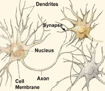 There are 100 Billion neurons in the brain. A typical neuron has about 1,000 to 10,000 synapses (that is, it communicates with many
other neurons, muscle cells, glands, etc.).
It has been estimated that there are 1 quadrillion synapses in the human brain. That's 1015. see neurons below.
There are 100 Billion neurons in the brain. A typical neuron has about 1,000 to 10,000 synapses (that is, it communicates with many
other neurons, muscle cells, glands, etc.).
It has been estimated that there are 1 quadrillion synapses in the human brain. That's 1015. see neurons below.This wiring system surpasses by many orders of magnitude the complexity of even the most advanced supercomputers.
In the 4th century BC Hippocrates concludes the brain was involved in sensation and was the seat of intelligence. Plato (428BC - 348BC) agrees, but it not accepted until experiments by Roman physiologist and anatomist Galen identified it in the 2nd century AD. See Neuroscience History But there is still a lot that is unknown. The brain is divided into the left and right hemispheres and with the exception of the pineal gland in the center, every brain module below is duplicated in each hemisphere.
So, a stroke in the left hemisphere may cause speech problems and paralysis of the right side of the body. Whereas a stroke in the right hemisphere may cause paralysis to the left side and problems with spatial and perceptual abilities causing misjudgement of distances and instability. See Effects of Stroke. The main components of the brain are most apparent in the embryonic development of the brain, where three swellings occur for the forebrain, midbrain and hindbrain. All vertibrates have these 3 parts.
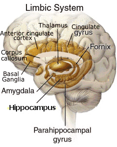
Simplified version: Limbic System - Emotion (feelings, relationship/nurturing, images and dreams, play), shared by mammals. Reptilian Brain - Instinct (survival, breathing/swallowing/heartbeat, startle response), entire brain in reptiles.
The following section is more about psychology than anatomy, but it is integrally linked with brain function.
White Matter - Gray Matter: Gray Matter - The real processing is concluded in the grey matter. They have shorter axons without a myelin sheath. | |
The Forebrain
* Telencephalon
|
 Source: Rice U. See Also: Functional brain map below |
|
The neopallium is the top layer of the cerebral hemispheres, about 2 mm thick, and is involved in higher functions such as sensory perception, generation of motor commands, spatial reasoning, and in humans, language and conscious thought. Other names for the neopallium include neocortex , isocortex and homotypical cortex. * Corpus Callosum - A bundle of nerve fibers (millions of axons from the cerebral cortex) that allows the two hemispheres to communicate. Cerebral Lobes: | |
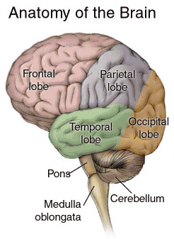 Source: Anatomy of the Brain at The American Health Assistance Foundation |
|
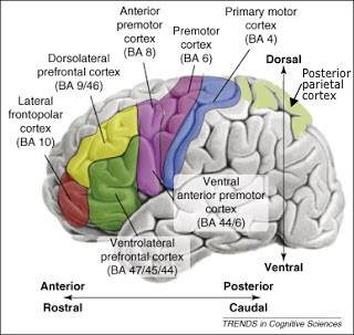 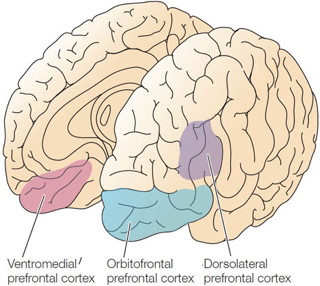 BA - Brodmann Area - Brodmann's map of cytoarchitectonics. See: The Human Brain | umich.edu/~cogneuro and Human brain - General features - Wikipedia The frontal lobe has 3 main division consisting of the prefrontal cortex and the pre-motor area and the motor area. The pre-motor and motor areas of the frontal lobes contain nerves that control execution of voluntary muscle movement and the prefrontal cortex is responsible for personality and expression and the planning of complex cognitive behaviour.. The Prefrontal cortex consists of the dorsolateral frontal cortex and the orbitofrontal cortex. Source: Cognitive Neuropsychology 101: Frontal Lobes !
Besides the frontal cortex, the posterior parietal cortex clearly plays a role in voluntary movements, by assessing the context in which they are being made. The parietal cortex receives somatosensory, proprioreceptive, and visual inputs, then uses them to determine such things as the positions of the body and the target in space. It thereby produces internal models of the movement to be made, prior to the involvement of the premotor and motor cortices.
See The hindbrain for the Medulla, Pons and Cerebellum. | ||||||||||||||||||||||
| ||||||||||||||||||||||
|
| |||||||||||||||||||||
* Diencephalon - Inner Brain - Part of Forebrain next to Midbrain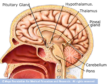
| ||||||||||||||||||||||
Brainstem- The brainstem connects the brain and the spinal cord. It consists of the medulla (an enlarged portion of the upper spinal cord), pons and midbrain (lower animals have only a medulla). The brainstem controls the reflexes and automatic functions (heart rate, blood pressure), limb movements and visceral functions (digestion, urination).The Midbrain (mesencephalon)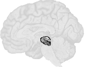 The midbrain (mesencephalon) occupies only a small region in humans (it is relatively much larger in "lower" vertebrates).
The Hindbrain (rhombencephalon)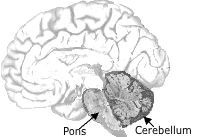 Cranial Nerves
Vagus Nerve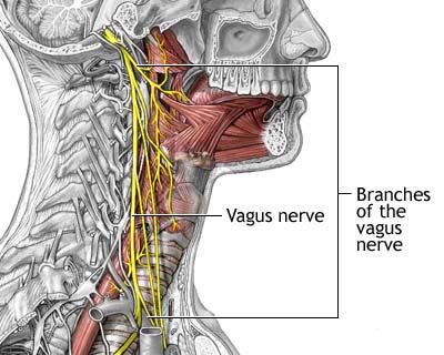 The vagus nerve is the longest of the cranial nerve. Its name is derived from Latin meaning "wandering". It does not go thru the spinal cord, but from the medula (part of the brainstem) down the neck, to the chest and abdomen.
 The pharyngeal branch travels between the internal and external carotid arteries and enters the pharynx at the upper border of the middle constrictor muscle. It supplies the all the muscles of the pharynx and soft palate except the stylopharyngeas and tensor palati. The superior laryngeal nerve branches distal to the pharyngeal branch and descends lateral to the pharynx. The third branch is the recurrent branch of the vagus nerve and it travels a different path on the left and right sides of the body. Speech is permitted through a branch of the vagus nerve, the laryngeal nerve, which innervates the larynx. In the thorax branches go to the lungs for bronchoconstriction, the esophagus for peristalsis and the heart for slowing of heart rate. In the abdomen branches enter the stomach, pancreas, small intestine, large intestine and colon for secretion and constriction of smooth muscle. See Vagus nerves at Medical Look Human Anaatomy Vagus nerve in health. | ||||||||||||||||||||||
Brain Map: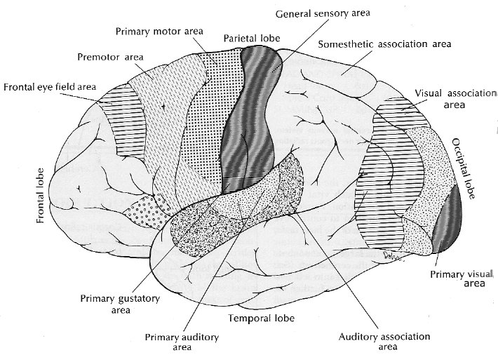 Source: The Columbia College of Physicians and Surgeons Complete Home Medical Guide See also the Functional brain map from Science Daily
Other Maps:
Sources:
A new brain atlas and database developed by the International Consortium for Brain Mapping (ICBM), uses an anatomically labeled brain template. The Laboratory of Neuro Imaging (LONI) at UCLA has a Data Immersive Visualization Environment (DIVE) for Brain Visualization. Brain WavesThe brain functions by sending electrical signals from one place to another. Very small charges pass between nerve cells, accompanied by changes in electrical potential, in voltage. This activity can be measured and displayed as a wave form called brain wave or brain rhythm.Brain waves are measured with an electroencephalogram (EEG) by placing electrodes on the scalp and measuring voltage differences.
NeuronsThe brain and spinal cord are made up of many cells, including neurons and glial cells.90% of the brain is glial cells; they provide support functions for the neurons. 
There are 100 Billion neurons in the brain. The neuron body contains organelles such as mitochondria, endoplasmic reticulum, and a nucleus (which contains DNA). The dendrites radiate out from the cell body. They are specialized for receiving information from other neurons. Dendrites act like antennae for nerve cells. The axon generally carries nerve impulses away from the cell body. Usually only one axon sprouts off of the cell body, but it may have many branches conecting to dendrites on many other neurons.. Axons convey information from one part of the brain to antoher. They may be up to a meter long.At birth, a babty already has about all of the neurons it will ever have, but it will continue to develop synapses as it learns throughout it's life. . It's brain doubles in size in the first year, and by age three it has reached 80 percent of its adult volume. Even more importantly, synapses are formed at a faster rate during these years than at any other time. In fact, the brain creates many more of them than it needs: at age two or three, the brain has up to twice as many synapses as it will have in adulthoo. These surplus connections are gradually eliminated throughout childhood and adolescence, a process sometimes referred to as blooming and pruning. Baby's Brain Begins Now: Conception to Age 3 | Urban Child Institute
Different Types Of Neurons
 Source: NIH Note: Some articles state that appendages of a sensory neuron that comes from the nerve ending is a dendrite. Women have 10% more neurons. You loose 10% over a lifetime. A typical neuron is 10 microns (µm) (.01 mm) in diameter. (See size) They vary in size from 4 microns (.004 mm) to 100 microns (.1 mm) in diameter. Their length varies from a fraction of an inch to several feet. The cell body of a motor neuron is approximately 100 microns (0.1 millimeter) in diameter.Neurotransmission Source: Rn Continuing Education When a dendrite is stimulated in a certain way, the neuron to which it is attached suddenly changes its electrical polarity and may fire, sending a signal out along its single axon where it may be picked up by the dendrites of other neurons.
A typical neuron has about 1,000 to 10,000 synapses (that is, it communicates with 1,000-10,000
other neurons, muscle cells, glands, etc.).
It has been estimated that there are 1 quadrillion synapses (the gap between the axon terminal
and the receiving cell) in the human brain. That's 1015.
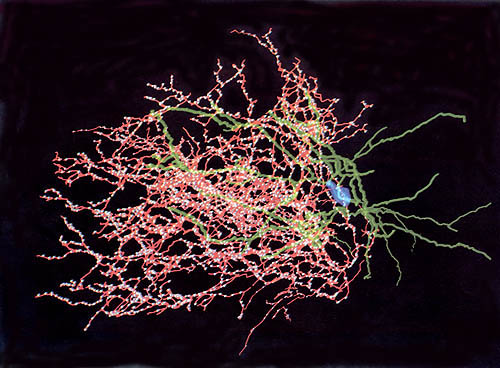
A computer model constructed
from an actual 3-d microscopic photo of a single cortical neuron. Infant brain development: At birth, a baby’s brain contains 100 billion neurons, and almost all the neurons the brain will ever have. A 2011 article says, At birth, the number of synapses per neuron is 2,500, but by age two or three, it’s about 15,000 per neuron. More synapses are created and some that are seldom or never used are eliminated.
New research says
New research in 2017 says there are over 1 million new connections per second in the first few years of life. Spinal Cord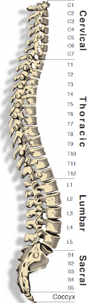 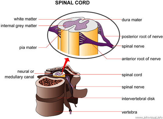
See other diagrams at: Spine under health,
Books:
See Also: Return to NeuroScience
| ||||||||||||||||||||||

 (A Giraffe's primary afferent axon (from toe to neck) is 15 feet long.)
The Synapse is the point of connection between two neurons (axon
and dendrite) or between a neuron (axon) and a muscle or gland.
Boutons are where synapes occur.
(A Giraffe's primary afferent axon (from toe to neck) is 15 feet long.)
The Synapse is the point of connection between two neurons (axon
and dendrite) or between a neuron (axon) and a muscle or gland.
Boutons are where synapes occur.